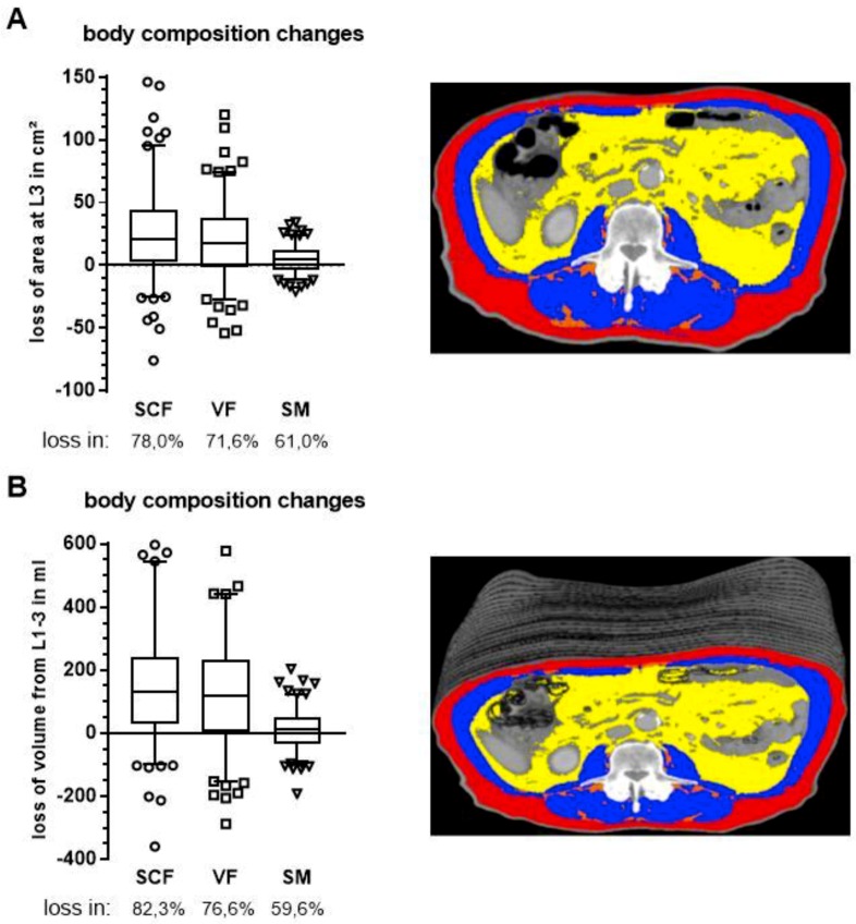
Figure 2.
Changes in body composition during CT for radiation planning and CT for first FU as determined by segmentation of subcutaneous fat (SCF, red), visceral fat (VF, yellow) and skeletal muscle (SM, blue) in a single CT slice at third lumbar vertebra (A) and as volumes (B) of stacked CT slices from the first to third lumbar vertebra. Respective patient images appear on the right. Boxes extend from 25th to 75th percentiles and whiskers are drawn down to 5th percentiles and up to the 95th percentiles.

