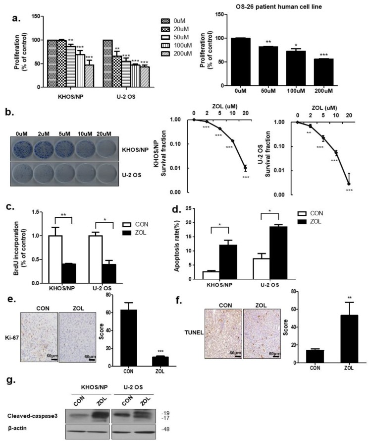
Figure 1.
Zoledronic acid (ZOL) inhibited osteosarcoma (OS) cell proliferation. (a) Cell viability was evaluated by MTT assay in KHOS/NP cells, U2OS cells of OS cell lines and cells from a patient with OS after 48 h treatment with the indicated concentration of ZOL; * p < 0.05, ** p < 0.01, *** p < 0.001. (b) Colony-formation assays were performed using KHOS/NP and U2OS cells treated with the indicated concentration of ZOL for seven days; ** p < 0.01, *** p < 0.001. (c) Cells were treated with ZOL (40 μM) for 72 h, and the proliferation rate was detected by 5-bromo-2′-deoxyuridine (BrdU) labeling; * p < 0.05, ** p < 0.01. (d) The apoptosis rate was assessed by fluorescence-activated cell sorting (FACS) analysis for 72 h treatment; * p < 0.05. (e) Ki67 expression in the orthotopic model was examined by immunohistochemistry; *** p < 0.001. (f) Two weeks after tumor cell inoculation, mice were randomly assigned into four groups of three animals each: Control group (untreated), ZOL alone group. ZOL was administered intraperitoneally twice weekly at a dose of 0.1 mg/kg in 100 μL PBS two weeks after inoculation. TUNEL assays were performed using orthotopic cells [23]; ** p < 0.01. (g) Immunoblotted cell lysates (30 μg) are shown with the cleaved caspase3 and β-actin antibodies for 48 h treatment.

