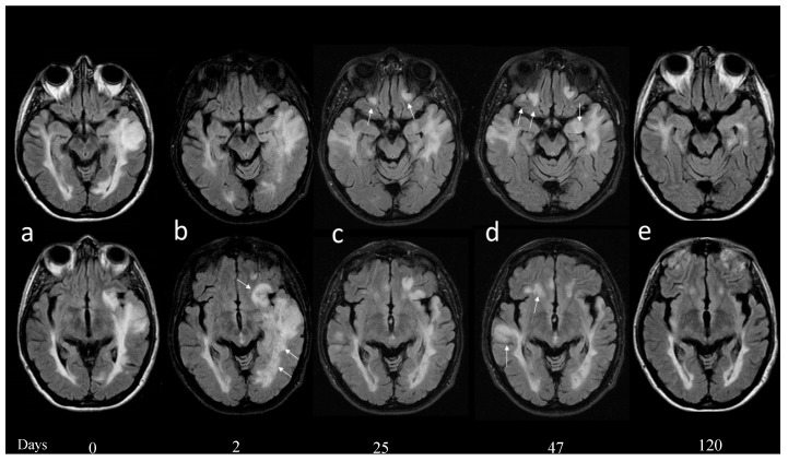
Figure 1.
At admission (a) showed a large T2 signal alteration involving the left temporal lobe and expanding the superior temporal gyrus. (b) Two days later, a new brain magnetic resonance imaging (MRI) showed increased edema also involving the frontal subcortical and periventricular white matter (b, arrows); compression of the ventricular system was increased; and no diffusion restriction or contrast enhancement was demonstrated. (c) Twenty-five days after admission a new MRI showed reduction of the previously described T2 temporal lobe signal alteration with reduced compression of the temporal horn of the ventricle. Five new lesions were demonstrated and two of these were located in the fronto-orbital regions bilaterally (c, arrows). (d) Forty-seven days after admission, a new MRI documented a worsening of the T2 signal alterations in the fronto-orbital and in the temporal region on the right, with mild contrast enhancement in the right hippocampus and cingulum cortex (not shown). (e) Four months after the beginning of symptomatology all the signal alterations were markedly reduced and no enhancement was evident.

