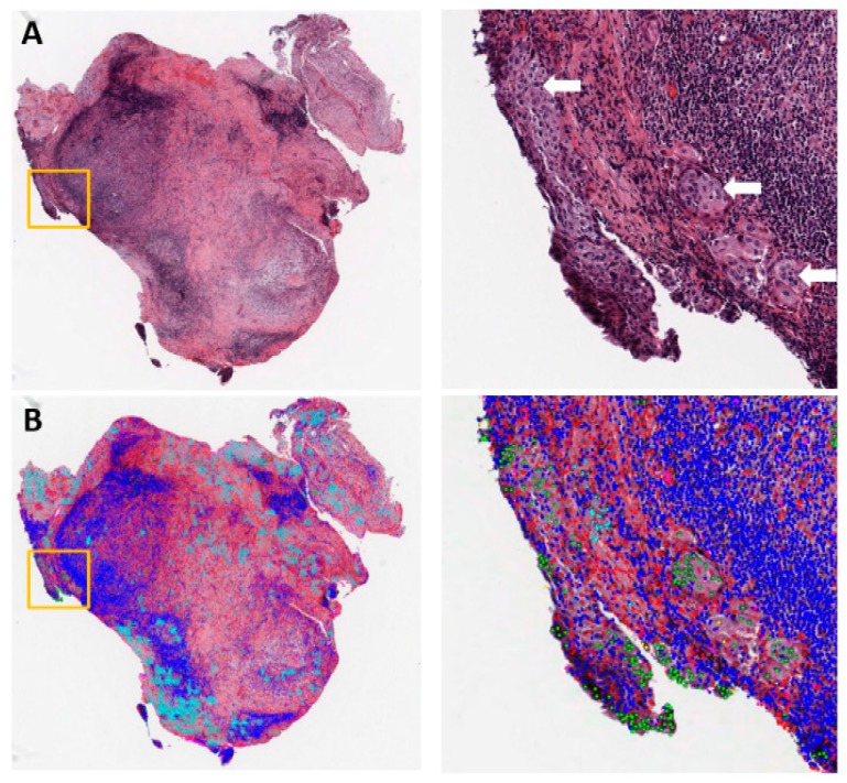
Figure 1.
Example of metastasis detection in the lymph node for a lung adenocarcinoma patient. (A) Left: Hematoxylin and eosin (H&E)-stained lymph node pathology slide (40×). Data were collected by the National Lung Screening Trial (NLST). Tumor cells began to invade into the capsule in the orange box. Right: Region of interest in the orange box on the left, with white arrows pointing to tumor cells. (B) Cell classification result overlaid on the H&E image. Green: Tumor nuclei; blue: Lymphocytes; red: Stroma nuclei; cyan: Necrosis.

