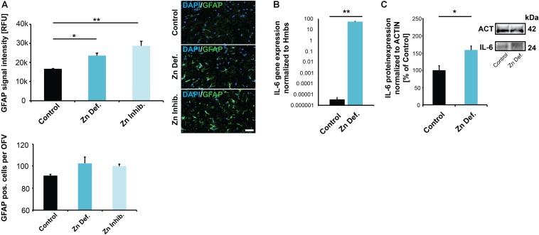
FIGURE 4.
(A) Brain sections from three animals per group were used for immunohistochemistry. DAPI (labeling cell nuclei) and GFAP was visualized and fluorescent signal intensities from 10 cells in the hippocampus from three sections per animal measured. Mean values show the average of three animals per group. Upper panel: A significant increase in GFAP expression levels can be detected in the brains of mice subjected to zinc deficiency and lowered bioavailability (Zn Inhibitor) compared to control mice. Lower panel: No significant difference in the number of GFAP positive cells per optic field of view (OFV) was found. Scale bar = 100 μm. (B) Whole-brain total RNA lysate from three animals per group was used to analyze the expression of IL-6 on gene level normalized to Hmbs. A significantly higher IL-6 expression was found in the brain of zinc-deficient mice. (C) Whole-brain protein lysate from three animals per group was used to analyze IL-6 expression on protein level normalized to ACTIN. Significantly higher IL-6 levels are found in the brain of zinc-deficient mice.

