Figure 1.
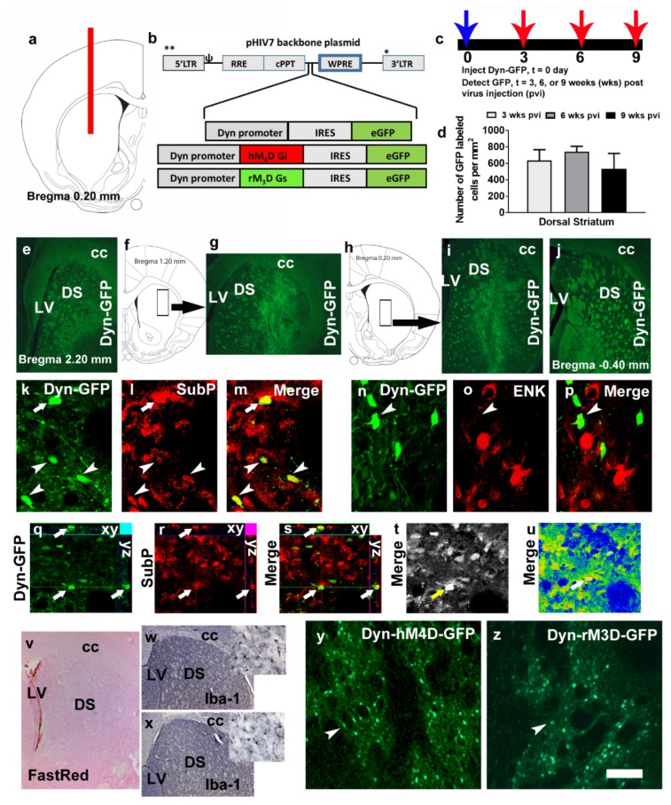
(a) Schematic representation of a coronal section through the dorsal striatum of the adult rat brain indicating the placement of injector needle for virus infusions. (b) Schematic of the lentiviral vector backbone indicating the genes of interest along with the dynorphin (Dyn) promoter that are inserted upstream of the WPRE in the pHIV-7 vector; IRES, internal ribosome entry site; eGFP, enhanced green fluorescent protein. (c,d) Time course of Dyn-GFP virus infection demonstrated that maximal expression was seen between 3–6 weeks after virus injection. (d) Quantitative analysis of Dyn-GFP positive cells in the striatum of virus injected animals; n = 2–4 each time point. Data is represented as mean ± SEM. (e–j) Coronal sections with Dyn-GFP positive neurons along the rostral-caudal direction of the dorsal striatum. The rectangular box in f, h indicates the striatal area labeled with Dyn-GFP neurons in g, i. LV, lateral ventricle; DS, dorsal striatum; cc, corpus callosum. (k–m) Colabeling of Dyn-GFP with SubP (CY3, red) a maker for D1R-MSNs; arrow and arrowheads in k–m point to colabeled immunoreactive cells. (n–p) Colabeling of Dyn-GFP with ENK (CY3, red) a maker for D2R-MSNs; arrowhead in n–p point to Dyn-GFP cell that is not colabeled with ENK cells. (q–s) Confocal z-stack images in orthogonal view indicating colabeling of the cell in (k–m) pointed with an arrow. Xy- and yz axis is indicated in q-s to demonstrate equal penetration of GFP and SubP antibodies. (t,u) Confocal images indicating detector gain (t; black and white image shows no red—overmodulation or green—undermodulation of cells and therefore the lasers have been optimized in the multi-channel image acquisition) and amplifier gain (u; rainbow image shows no red—overmodulation or blue—undermodulation of areas expressing cells; note that the area of the axon bundles are blue due to lack of any cellular bodies) of the section used for orthogonal view. GFP, green fluorescent protein; SubP, substance P; ENK, enkephalin. (v) Virus injected section stained with Vector FastRed showing minimal damage to the dorsal striatum. (w,x) Iba-1 staining via DAB in virus naïve (w) and Dyn-GFP (x) injected rat. (y,z) GFP immunoreactivity in the dorsal striatum of a rat injected with Dyn-hM4D-GFP (y) and Dyn-rM3D-GFP (z). Scale bar in u is 200 um applies to e, g, i, j, v, w, x; 20 um applies k–p; 30 um applies q–u; 70 um applies y–z.

