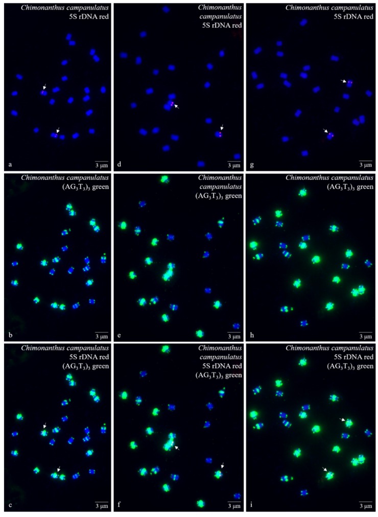
Figure 1.
Mitotic metaphase (preliminary stage) chromosomes of Chimonanthus campanulatus R.H. Chang & C.S. Ding after fluorescence in situ hybridization (FISH). Three spreads are presented in Figure 1a–c, Figure 1d–f, and Figure 1g–i. The probe oligo–5S rDNA result with chromosomes visualized by carboxytetramethylrhodamine (TAMRA) (red, white arrows) is shown in Figure 1a,c,d,f,g,i, whereas the probe oligo–(AG3T3)3 result with chromosomes visualized by carboxyfluorescein (FAM) (green) is shown in Figure 1b,c,e,f,h,i. Chromosomes were counterstained by 4,6-diamidino-2-phenylindole (DAPI) (blue) in all images. Scale bar = 3 μM.

