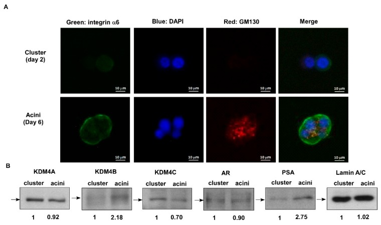
Figure 1.
Decrease of human histone demethylase (KDM)4C expression during acinar morphogenesis of human RWPE-1 prostate epithelial cells. (A) Confocal images of RWPE-1 cells forming prostatic organoids in three-dimensional (3D) culture. The structures were immune-stained with basal extracellular membrane receptor α6-integrin (green) and the apical marker GM130 (red). Nuclei were counterstained with DAPI dye (blue). Scale bar = 10 μm. The RWPE-1 cells in 3D culture were collected on the 2nd day for cluster morphology and on the 6th day for acinar morphology for further protein expression analysis. (B) Protein expression of KDM4A, KDM4B, KDM4C, androgen receptor (AR), and prostate specific antigen (PSA) in cluster versus acinar morphology of RWPE-1 cells was determined by Western blotting. Lamin A/C was used as loading control.

