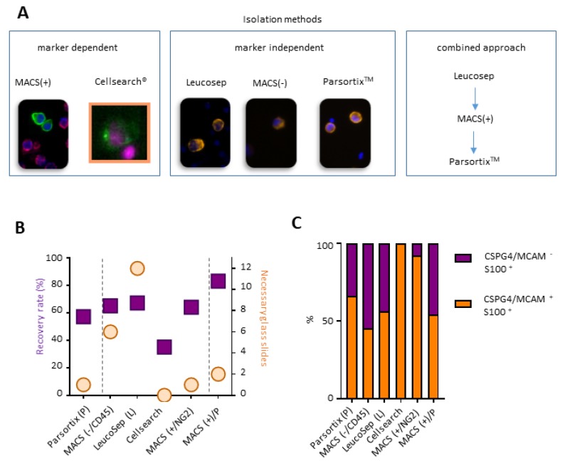
Figure 2.
Method to analyze melanoma cell subsets. (A) Tested isolation methods for the membrane marker dependent and independent enrichment of melanoma cells. (B) Recovery rate of 50 melanoma cells in 7.5 mL of blood and subsequent histological slides needed for analysis. (C) Amount of melanoma cells positive for membrane markers CSPG4/MCAM and intracellular S100. N = 3–4.

