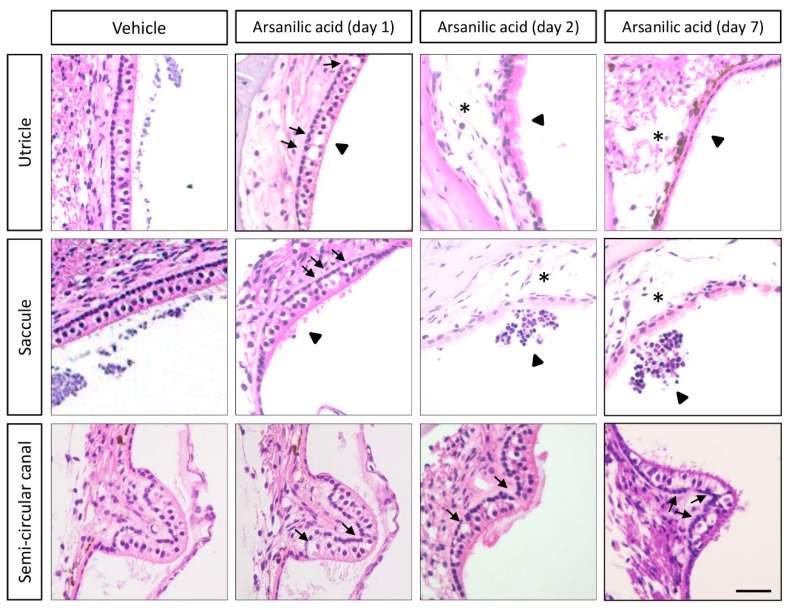
Figure 2.
Histological observations (hematoxylin–eosin staining) after unilateral labyrinthectomy with arsanilic acid or phosphate buffer (vehicle) in mice. The vehicle group shows negligible damage in the utricles, saccules, and semi-circular canals. In the arsanilic acid group, vacuoles can be seen in the utricles and saccules at 1 day after surgery (arrows). The vacuoles gradually increase in size, and the macular structures are destroyed by 2 days after surgery (asterisk). The vacuoles are also observed in the semi-circular canals at 1, 2, and 7 days after surgery (arrows). In the utricles and saccules, the disarrangement and loss of otoconia could be observed (arrowheads). Scale bar, 20 μm.

