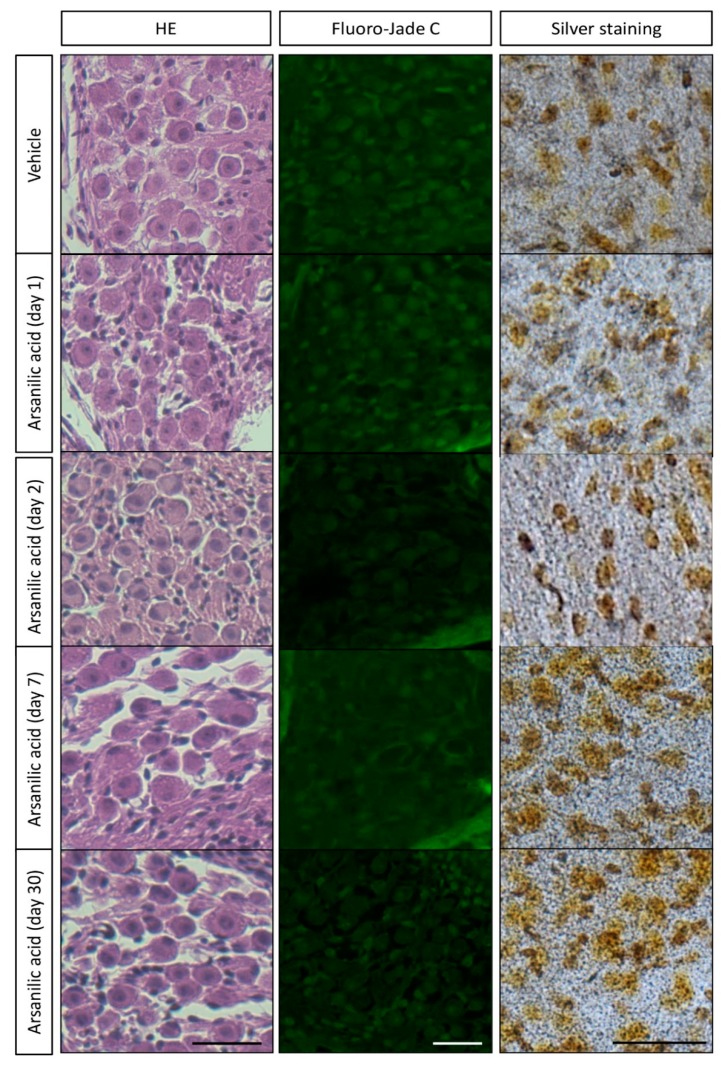
Figure 3.
Observation of cells in mouse vestibular ganglions after staining with hematoxylin–eosin, Fluoro-Jade C, and silver. Paraffin sections of temporal bone were stained with hematoxylin–eosin (HE) and Fluoro-Jade C to check cellular integrity and degeneration. We further employed an amino-cupric silver staining method that is very sensitive in detection of cellular degeneration. For the silver staining, we used both frozen sections and vibratome sections of the vestibular ganglions. Because the frozen sections were of uniform thickness and gave us clearer images, representative pictures of frozen sections are shown here. There is no difference in the amount of structural damage observed with hematoxylin–eosin staining, or in the number of cells stained by Fluoro-Jade C and silver, between the arsanilic acid (unilateral labyrinthectomy with arsanilic acid) and vehicle (unilateral labyrinthectomy with phosphate buffer) groups. Scale bar, 20 μm.

