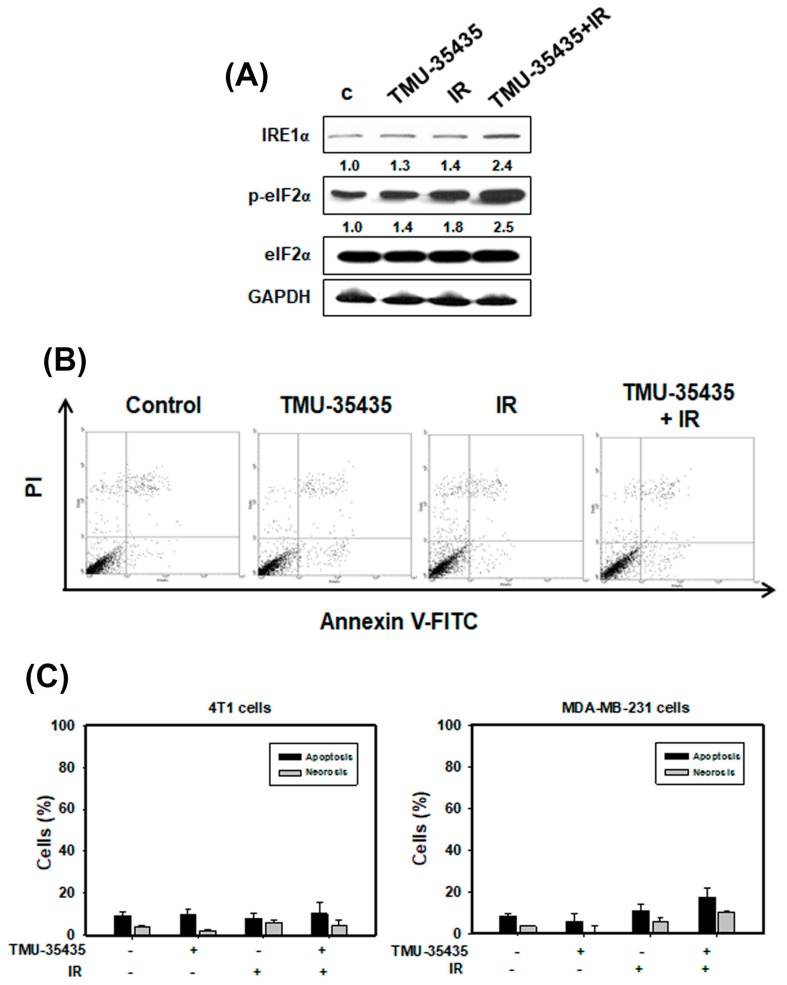
Figure 3.
Measurement of endoplasmic reticulum (ER) stress and apoptosis in cells treated with different groups. (A) Effects of TMU-35435 and IR on the expression of ER stress-associated proteins. The cells were treated with IR (4 Gy) and TMU-35435 (1 μM) for 12 h. (B) Apoptosis was analyzed by an Annexin V apoptosis kit using flow cytometry. The cells were treated with TMU-35435 (1 μM) and IR (4 Gy) for 24 h. (C) Quantification of apoptosis and necrosis in MDA-MB-231 and 4T1 cells that received various treatments. The cells were treated with TMU-35435 (1 μM) and IR (4 Gy) for 24 h.

