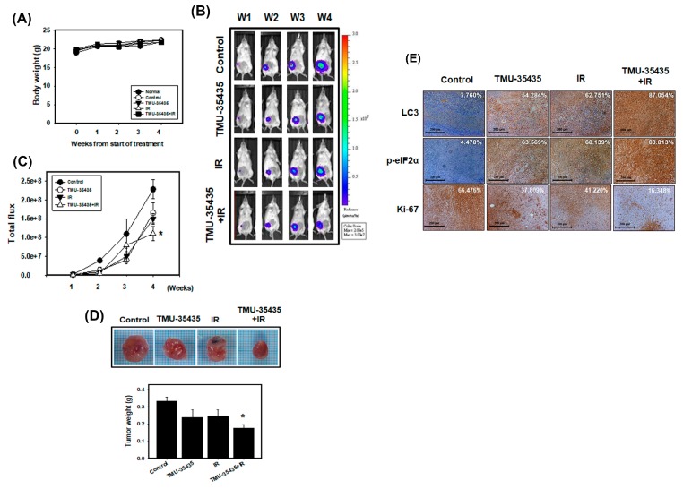
Figure 7.
Combined treatment with TMU-35435 and IR increases antitumor effects in a mouse model of orthotopic breast cancer. (A) Measurement of body weight in Balb/c mice was evaluated once per week. (B) 4T1-Luc cells were injected into Balb/c mice of the mammary fat pads, which were analyzed for luciferase signals by an in vivo imaging system (IVIS) 200. (C) Quantification of the luciferase signals. (D) The tumor weight was measured in the Balb/c mice after sacrifice. * p < 0.05 versus control. (E) Immunohistochemistry (IHC) staining of orthotopic tumor tissues. The LC3 and p-eIF2α expression was determined by IHC staining. The percentage of positive cells was analyzed by HistoQuest software (TissueGnostics). Scale Bar: 200 μm.

