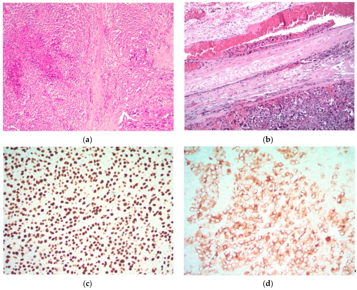
Figure 1.
Parathyroid carcinoma is characterized by sheets of parathyroid cells with fibrosis and focal necrosis (top left). The diagnosis is confirmed by the identification of unequivocal vascular invasion defined as tumor cells within vascular channels associated with fibrin (top right). The tumor cells exhibit nuclear positivity for GATA3 (bottom left) and cytoplasmic positivity for chromogranin (bottom right) and parathyroid hormone (not shown), confirming parathyroid differentiation. Original magnification (a) ×120; (b) ×200; (c) and (d) ×400.

