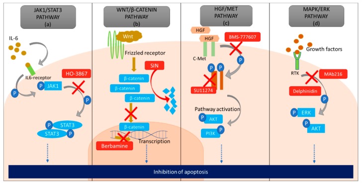
Figure 4.
A diagrammatic representation of the pathways that can indirectly lead to inhibition of apoptosis in a cancer cell. (a) Janus activated kinase 1 (JAK1)/signal transducer and activation of transcription-3 (STAT3) pathway is upregulated due to the accumulation of interleukin 6 (IL-6) which causes phosphorylation of JAK1 and STAT3, promoting cell survival and increase proliferation. However, HO3867 inhibits phosphorylation of STAT3 causing apoptosis. (b) Wnt pathway is activated by the binding of Wnt molecule to the frizzled receptor (FZD). This causes the formation of a complex and accumulation of β-catenin in the cytoplasm. Saturation of β-catenin in cancer cells causes its translocation into the nucleus, where it initiates uninterrupted transcription leading to an increase in proliferation and chemoresistance. However, sinomenine (SIN) degrades β-catenin in the cytoplasm and berbamine inhibits transcription in the nucleus of cancer cells eventually causing apoptosis. (c) Hepatocyte growth factor (HGF) pathway initiates upon binding of HGF to c-Met (tyrosine kinase receptor) which leads to phosphorylation of c-Met, which in turn activates various other cell signaling pathways including AKT, PI3K pathways important for cell survival, proliferation and angiogenesis. BMS-777607 obstructs the phosphorylation of c-MET and SU11274 decreases the concentration of c-MET in the cell to inhibit HGF pathway, both leading to apoptosis (d) Growth factors attach to receptor tyrosine kinase (RTK) present on the extracellular surface of the cell and can switch on the mitogen-activated protein kinase (MAPK)/extracellular signal-regulated kinase (ERK) pathway in the cell. An increased concentration of growth factors leads to over-activation of MAPK/ERK pathway in cancer cells. Delphinidin and Mab216 down-regulates this pathway by inhibiting phosphorylation of ERK, AKT, which eventually leads to apoptosis.

