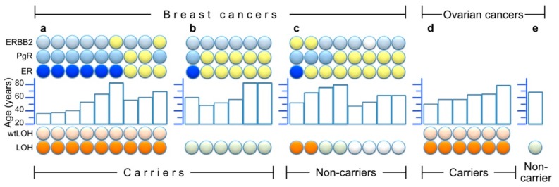
Figure 4.
Summary of LOH results and/or histological staining of estrogen-, progesterone- and HER2/Erb-B2 receptors (ER, PgR and ERBB2, respectively) in 30 tumors. Within each partition of the figure, the samples are ordered from left to right by LOH results (bottom symbols: orange for LOH and light orange above that, denoting wtLOH, light green for samples without LOH, and white for unavailable LOH information), and then by ER results (ER line of symbols: blue for ER negative, yellow for ER positive) and finally by age (middle histogram) and other receptor results (top two lines of symbols: light blue = negative, yellow = positive and white = unavailable). (a) Nine breast cancers from heterozygous carriers who had wtLOH in their tumors; (b) Six breast cancers from heterozygous carriers without LOH; (c) Eight breast cancers from non-carriers among close (1st and 2nd degree) relatives of genotyped or obligate carriers (four of which were available for LOH analysis of microsatellite markers); (d) Six ovarian cancers from carriers; (e) One ovarian cancer from a non-carrier.

