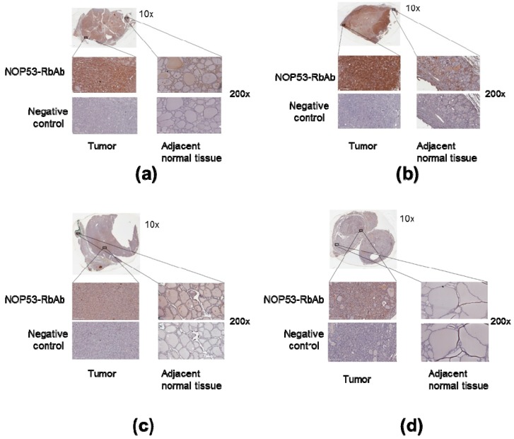
Figure 6.
Overexpression of NOP53 in tumors from patients with FNMTC. Panels A through D show representative immunohistochemical staining for NOP53 in thyroid cancer samples from the four affected members of Kindred 2: (a) Corresponds to Patient III.1.; (b) Patient II.2.; (c) Patient III.2.; and (d) Patient II.1. Each panel contains an inlet (zoom 10×); two separate regions—from tumor tissue and adjacent normal thyroid tissue—of higher magnification images (zoom 200×); and two higher magnification images (zoom 200×) from a negative control specimen at a similar location. The top left represents tumor staining with NOP53-RbAb, the top right shows adjacent normal tissue staining with NOP53-RbAb, and the two bottom images are negative controls. We observed that the tumor tissue showed a higher expression of NOP53 compared to the adjacent normal thyroid tissue in the four patients studied.

