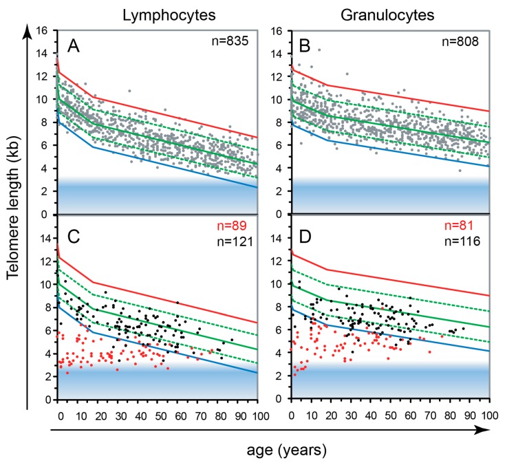
Figure 2.
(A,B) Telomeres in human lymphocytes and granulocytes from peripheral blood shorten with age [15]. Fluorescence in situ hybridization followed by flow cytometry (Flow FISH) results from over 800 healthy individuals were used to calculate the distribution of telomere length at any given age (percentiles in population: solid green 50th; red 1st, blue 99th). Note the marked variation on average, cell specific telomere length at any given age. (C,D) The critical role of telomerase is illustrated by the telomere length in cells from patients that are haplo-insufficient for telomerase genes (red dots in C and D) compared to their unaffected siblings (black dots in C and D). The blue zone at the bottom of the graphs represents the area where telomeres are expected to be fully “uncapped” with less than 1 kb of TTAGGG repeats per chromosome end. Reproduced with permission from [12].

