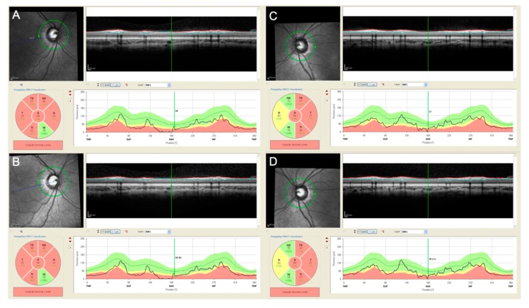
Figure 2.
Peripapillary retinal nerve fiber layer (pRNFL) analysis performed in both eyes (A–B right eye, C–D, left eye) with spectral domain optical coherence tomography (SD-OCT, HRA+OCT Spectralis, Heidelberg Engineering, Heidelberg, Germany) in a 4 years old patient affected by neurofibromatosis type 1 (NF-1) and a optic chiasmatic-hypothalamic glioma. The patient was already treated with systemic chemotherapy and during the follow-up the visual acuity remained stable (0.52 logMAR in the right eye and 0.10 logMAR in the left eye). The pRNFL analysis performed in both eyes at the end of the chemotherapy (A,C) and 6 months after (B,D) confirm the stability of the pRNFL thickness during time.

