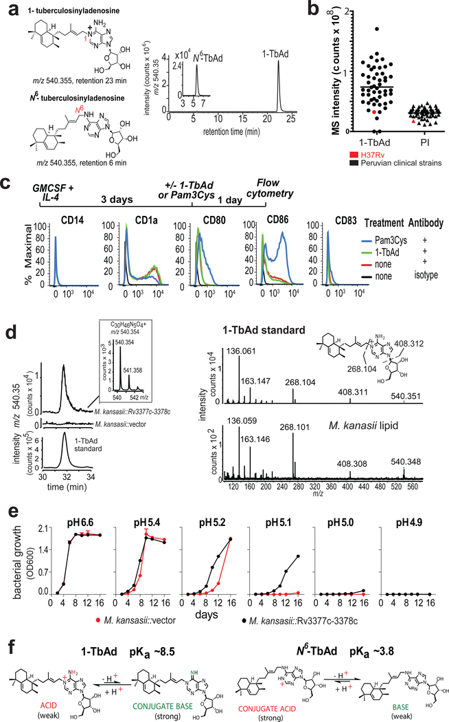
Figure 1. Testing signaling-dependent and signaling independent effects of TbAd on human cells.
a) 1-TbAd and N6-TbAd were detected as HPLC-MS ion-chromatograms. b) Raw MS counts for 1-TbAd and phosphatidylinositol (PI) were measured in 52 clinical strains and the laboratory strain H37Rv. c) Human monocytes were treated with cytokines followed by a TLR2 agonist (Pam3Cys) or 1-TbAd and subjected to flow measurement of activation markers in 3 independent experiments showing similar results. d) The Rv3377c-Rv3378c locus was transferred into M. kansasii, conferring production of a molecule with equivalent mass, retention and CIDMS fragments found in 1-TbAd. e) M. kansasii was grown in liquid media at various pH values in duplicate with low variance (coefficient of variation < 2 % for all but one measurement) in three independent experiments. f) pKa’s are derived from measurements of dimethylallyladenosine and related compounds(21, 22), which indicate that 1-TbAd is predominantly charged but in equilibrium with its uncharged conjugate base at neutral pH. Thus, 1-TbAd but not N6-TbAd exists as a strong conjugate base.

