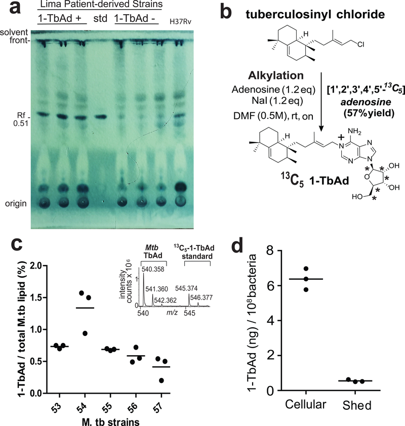
Figure 2. Quantitative measurements of 1-TbAd using an internal standard on a per cell basis.
a) Total lipids from three 1-TbAd+ and three 1-TbAd- strains were separated on normal phase silica TLC using chloroform/acetic acid/methanol/water mixtures in comparison to a 1-TbAd standard and a H37Rv lab strain. Lipids were subjected to charring after spraying with a phosphomolybdic acid solution. b) Labeled 1-TbAd was synthesized using 13C5 labeled adenosine whose mass spectral peaks did not overlap with those of natural 1-TbAd, allowing its use an in internal control. c) Total lipids from 5 patient-derived Mtb strains were weighed and subjected to HPLC-MS measurement in triplicate using the 13C-labeled internal standard to provide absolute mass values, which were expressed as the percentage of input lipid from 3 biologically independent samples.. d) For one representative clinical strain, lipids were extracted from the cell pellet (cellular) or conditioned media (shed) and measured as in c), expressed on a per cell basis based on colony forming unit (cfu) measurements from the input culture.

