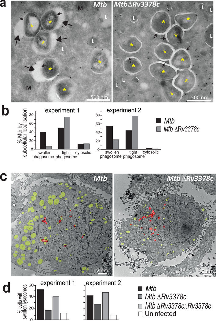
Figure 6. Infection of human macrophages for 4 days with Mtb or the Rv3378c-deletion mutant, which lacks TbAd production.
a) High magnification images of macrophage cytosol containing Mtb (*), mitochondria (M), lysosomes (L), capsular layer (small arrow) or intraphagosomal area with inclusions (large arrow). b) Greater than 150 intracellular Mtb were evaluated at high power for their localization in the cytosol, tight phagosomes (small arrows) or large phagosomes (p=7.0×10⁻¹⁵ for swollen versus tight phagosomes). c) Low magnification images show yellow pseudocoloring for swollen lysosomes (CD63 immunogold, electron lucent) and red pseudocoloring highlights individual Mtb bacilli. d) Greater than 150 cells were evaluated as having swollen lysosomes when one-third of the cytosolic area showed electron-lucent CD63+ compartments (p < 0.001 for Mtb versus MtbΔRv3378c; p < 0.05 for MtbΔRv3378c versus MtbΔRv3378c::Rv3378c). All p values in b and d were calculated with the Cochran-Mantel-Haenszel test with independent experiments treated as strata and were adjusted using the method of Benjamini and Hochberg.

