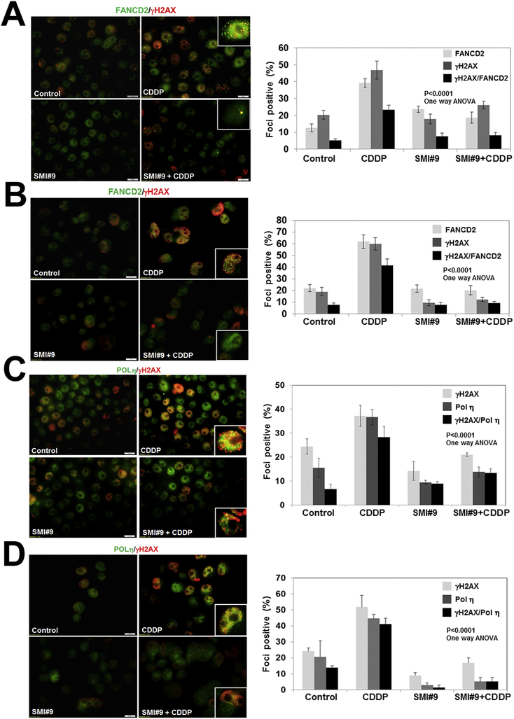
Fig. 6. SMI#9 treatment impedes cisplatin-induced recruitment and colocalization of DNA damage response proteins in TNBC cells.
Dual immunofluorescence staining of FANCD2/γH2AX, POL η/γH2AX, or RAD51/γH2AX in MDA-MB-468 (A,C,E) or SUM1315 (B,D,F) cells treated with 1 μM (SUM1315) or 3 μM (MDA-MB-468) CDDP with or without pretreatment with 1 μM SMI#9. Scale bars, 20 μm. Graphs show foci positive cells scored from at least 100 cells in five-ten fields from two independent experiments by Image J. Cells containing > 5 colocalized foci were counted for protein colocalization. Results were analyzed by 2-tailed Student’s t-test. Details of treatment conditions are provided under Methods.


