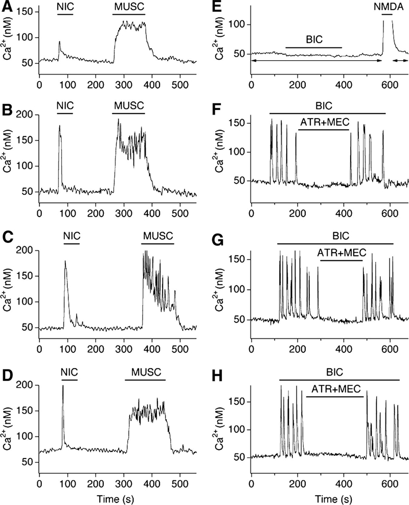
FIG. 4.
NMDAR1 knockout increases cholinergic responsiveness and cholinergic activity in mouse hypothalamic cultures. Representative Ca2+ imaging recordings from neurons in control (A, E), AP5 + CNQX–treated (B, F), AP5-treated (C, G), and NMDAR1 knockout (D, H) cultures are shown. A–D: traces illustrate neuronal responses to nicotine (NIC) and muscarine (MUSC). In A–D, tetrodotoxin (TTX, 2 μM) was in all media. E–H: traces illustrate ACh-dependent activity. Typical control neuron did not respond to bicuculline (BIC; 50 μM) in the presence of glutamate receptor antagonists (E). However, neurons in the 3 experimental groups revealed Ca2+ activity during disinhibition (F–H). The activity was cholinergic as was suppressed by AChR antagonists (ATR + MEC; 100 μM each). AP5 (100 μM) and CNQX (10 μM) were in all solutions in A–H except for the NMDA-containing (10 μM)/Mg2+-free solution in E used to confirm that the cells were healthy and responsive (the presence of AP5 + CNQX in E is shown by arrows).

