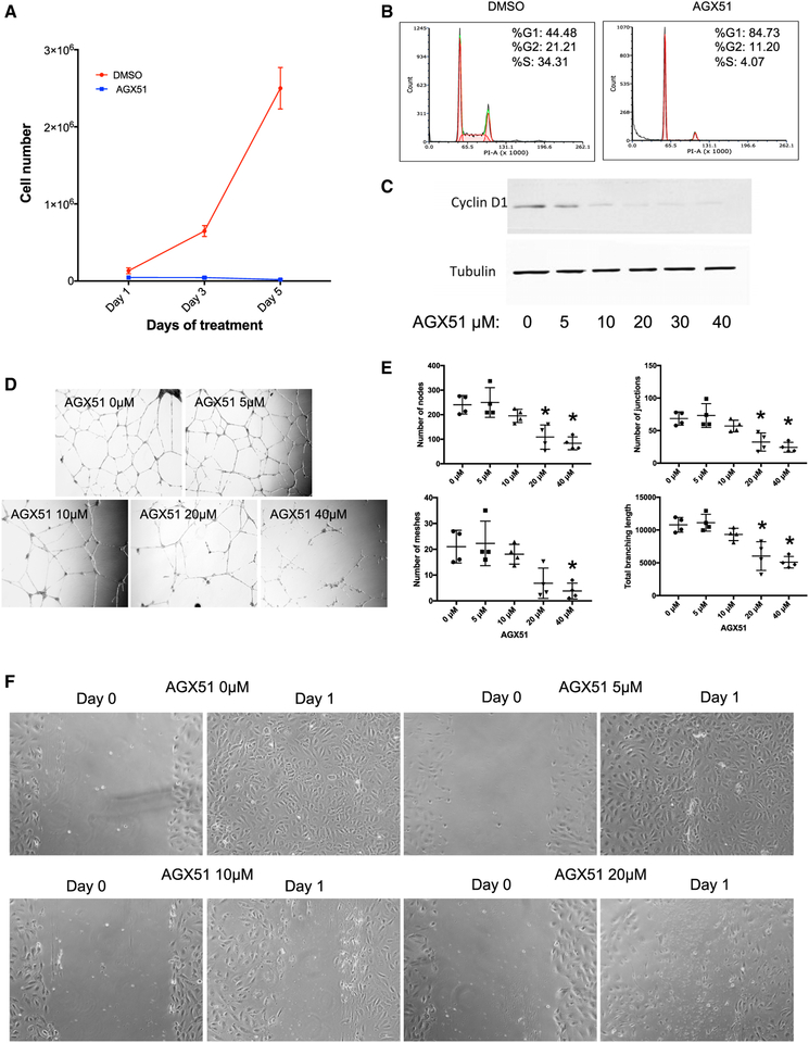
Figure 4. Effects of AGX51 on HUVEC Growth.
(A) Cell growth of HUVECs treated with DMSO or 20 μM AGX51 for 5 days.
(B) Cell cycle analysis of HUVECs treated with DMSO or 20 μM AGX51 for 24 h.
(C) Western blot for Cyclin D1 on whole-cell lysates from HUVECs treated with 0–40 μM AGX51 for 24 h. Tubulin is used as a protein loading control. See also Figure S5.
(D) HUVEC branching was observed after 18–20 h of culturing on matrigel in the absence or presence of 0–40 μM AGX51; images were taken at 10× magnification.
(E) Quantification of the number of nodes, junctions, meshes, and total branching length (n = 4 replicates per concentration tested); *p < 0.05 by Wilcoxon test.
(F) HUVEC monolayers were scratched, then media were replaced with media containing 0–40 μM AGX51, and migration was observed after 24 h, with images taken at 20× magnification.

