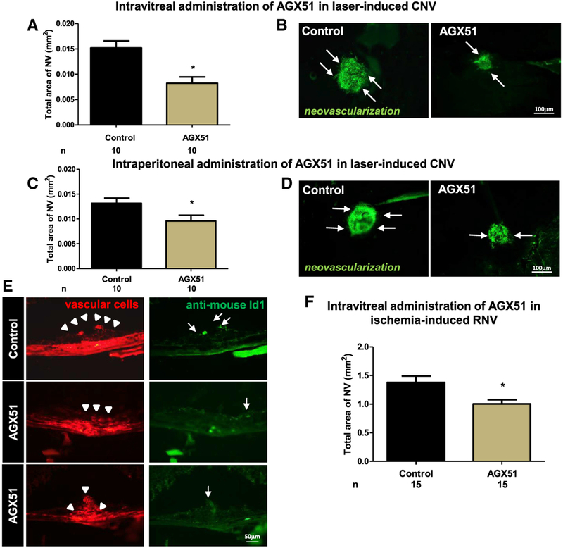
Figure 5. AGX51 Treatment Suppresses Ocular Neovascularization in Mouse Models of AMD and ROP.
(A) Wild-type mice had rupture of Bruch’s membrane at three locations in each eye, followed by intravitreal injection of 10 μg of AGX51 or vehicle in one eye immediately and after 7 days (n = 10 per group). Fourteen days after laser-induced rupture, the mean area of CNV was significantly less in AGX51-injected eyes than control eyes (*p < 0.05 by ANOVA with Bonferroni correction for multiple comparisons; error bars represent SEM).
(B) Representative Griffonia-simplicifolia-lectin (marking vascular cells)-stained choroidal flat mounts from a control-injected eye and an AGX51-injected eye (bar represents 100 μm).
(C) Twice-daily i.p. injection of 500 μg of AGX51 also significantly suppressed CNV (n = 10 mice per group; *p < 0.05 by unpaired t test; error bars represent SEM).
(D) Representative Griffonia-simplicifolia-lectin-stained choroidal flat mounts of eyes from mice treated with AGX51 (500 μg) or vehicle by twice-daily i.p. injection (bar represents 100 μm).
(E) Immunofluorescence for Id1 of CNV regions from mice treated with AGX51 or DMSO by intravitreal injection (bar represents 50 μm).
(F) Pups (n = 15) were placed in a 75% O2 chamber at P7 to induce ROP. At P12, the mice were returned to room air and received intravitreal injection of 10 μg of AGX51 in one eye or DMSO in the FE. On P17, the mice were euthanized, and the area of retinal neovascularization (RNV) was assessed. FE refers to “fellow eye” and is defined as the untreated eye in an animal in which both eyes received the laser treatment (*p < 0.01 by unpaired t test; error bars represent SEM).

