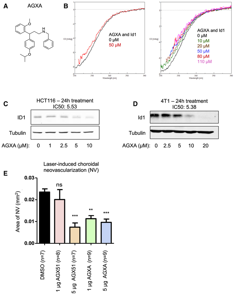
Figure 7. Characterization of the AGX51 Derivative AGXA.
(A) The chemical structure of AGXA.
(B) Circular dichroism (CD) spectra of AGXA (0–110 μM in DMSO) and Id1.
(C) Western blot for ID1 on whole-cell lysates from HCT116 cells treated with 0–10 μM AGXA for 24 h. The IC50 is indicated. The IC50 of AGX51 was 22.28 μM.
(D) Western blot for Id1 on whole-cell lysates from 4T1 cells treated with 0–20 μM AGXA for 24 h. The IC50 is indicated. The IC50 of AGX51 was 26.66 μM.
(E) Laser-induced choroidal neovascularization (NV) was induced in mice, and they were treated by intravitreal injection with DMSO, 1 or 5 μg AGXA, or 1 or 5 μg AGX51. On day 14, the animals were euthanized, and the area of CNV was measured (**p < 0.01, ***p < 0.0001 by ANOVA; error bars represent SEM).

