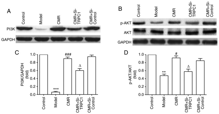
Figure 7.
Effect of CMR on PI3K/AKT signaling in the DRG neurons of diabetic rats. DRG neurons were separated and the lysates were analyzed using western blotting. (A) Expression of PI3K, (B) AKT and p-AKT proteins was detected by western blotting. (C) Relative expression of PI3K (PI3K/GAPDH) was statistically analyzed. (D) Relative expression of p-AKT (p-AKT/AKT) was statistically analyzed. GAPDH was used to confirm equal sample loading. **P<0.01, ***P<0.001 vs. control; #P<0.05, ###P<0.001 vs. model; ΔP<0.05 vs. CMR. CMR, Cortex Mori Radicis extract; TRPC1, transient receptor potential canonical channel 1; si-, small interfering RNA; DRG, dorsal root ganglia; P-, phosphorylated.

