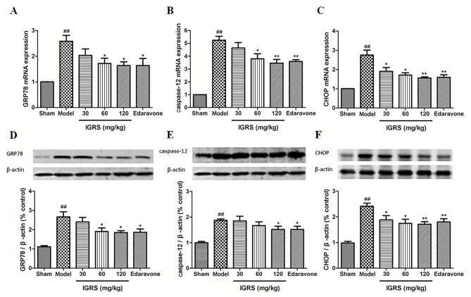
Figure 5.
Effect of IGRS on the expression of GRP78, caspase-12 and CHOP mRNA and protein in cerebral ischemia-reperfusion injured rats. The brain tissues of the experimental rats were removed, and the hippocampus tissues were separated from the injured side of the brain at 24 h post-reperfusion. The total RNA was extracted, cDNA was produced, and RT-qPCR was conducted. The expression of GAPDH was used as a loading control. The expression of β-actin was used as a loading control for western blot analysis. (A) GRP78, (B) caspase-12 and (C) CHOP mRNA levels were determined by RT-qPCR (n=5 per group). The expression levels of (D) GRP78, (E) caspase-12 and (F) CHOP protein were determined by western blot analysis (n=5 per group). Values are presented as mean ± standard deviation of each group. ##P<0.01 vs. Sham group. *P<0.05 and **P<0.01 vs. Model group. RT-qPCR, reverse transcription-quantitative polymerase chain reaction; IGRS, iridoid glycosides from Radix Scrophulariae; GRP78, endoplasmic reticulum chaperone BiP; CHOP, DNA damage-inducible transcript 3 protein.

