Figure 5.
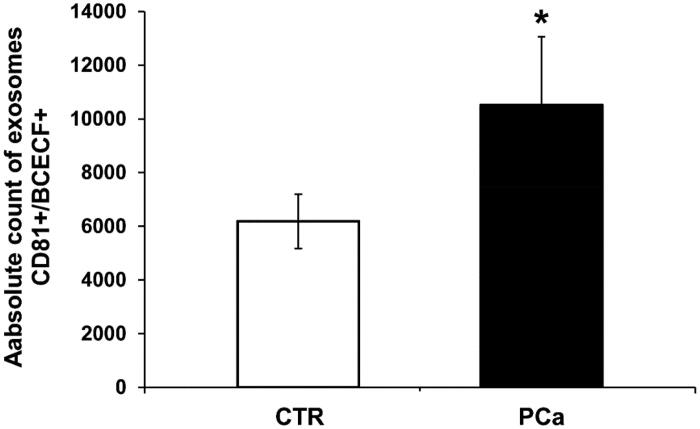
Nanoscale flow cytometry of plasma exosomes in PCa and CTR for intraluminal pH evaluation. The cytometer was calibrated using a mixture of non-fluorescent silica beads and fluorescent (green) latex beads with sizes from 110 nm to 1300 nm. The exosome preparation derived from plasma of 8 PCa patients and 8 CTR were stained 20 min at RT with anti-CD81 antibody and BCECF AM (10 µM) and analysed using flow cytometry. The double-positive events were then analysed for their size, based on the calibration with beads. Cumulative data are shown of the absolute number of CD81+/BCECF + exosomes of size less than 180 nm recovered from the plasma samples. Data are expressed as means ± SE. The p values was <.1 in PCa plasma exosomes compared to CTR. *p < .1.

