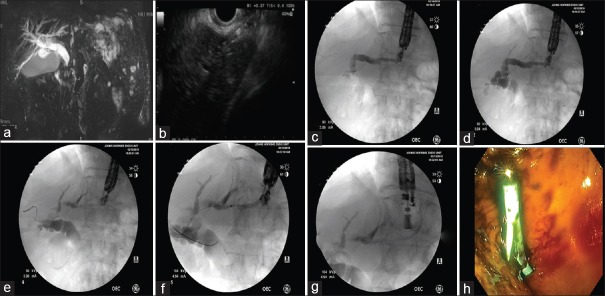
Figure 2.
EUS-hepatogastrostomy for malignant biliary obstruction in a 75-year-old male status postpartial gastrectomy and Roux-en-Y reconstruction for gastric cancer. (a) Significant dilatation of the common bile duct can be appreciated in the magnetic resonance imaging. (b) A linear echoendoscope was advanced to the stomach. Following the identification of the left intrahepatic duct, a 19-gauge needle was advanced transgastrically into the left intrahepatic duct. (c and d) Contrast was injected and anterograde cholangiography was performed, confirming correct positioning within the biliary tree. Dilatation of the intrahepatic and extrahepatic bile ducts can be appreciated. (e and f) A guidewire was passed into the left hepatic duct across the hilum into the common hepatic duct. (g) A 7 Fr × 10 cm plastic stent with internal and external flaps was placed over the wire into the left intrahepatic duct. (h) Endoscopic view of the deployed plastic stent

