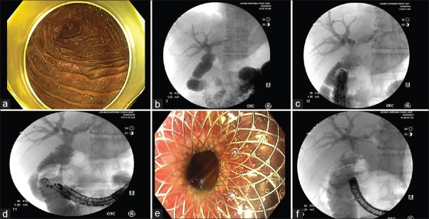
Figure 3.
EUS-directed transenteric ERCP for choledocholithiasis in a 50-year-old male with a history of Roux-en-Y hepaticojejunostomy due to bile duct injury postcholecystectomy. (a and b) The endoscope was advanced to the jejunum, and a mixture of contrast and saline was injected into the afferent limb, confirming correct position. (c) The endoscope was withdrawn, and a linear EUS was advanced to the duodenum and a location suitable for the duodenojejunal anastomosis was established under fluoroscopy. (d) The small bowel was punctured with a 19-G FNA needle. The small bowel adjacent to the hepaticojejunostomy anastomosis was dilated using 330 cc of saline mixed with contrast. Under EUS guidance, a cautery-assisted lumen-apposing metal stent, 15 mm × 10 mm, was then deployed creating the duodenojejunostomy. (e) Endoscopic view of the proximal flange of lumen-apposing metal stent post-deployment. (f) Under fluoroscopic guidance, using a therapeutic gastroscope, a guidewire was advanced across the lumen-apposing metal stent and hepaticojejunostomy was successfully cannulated

