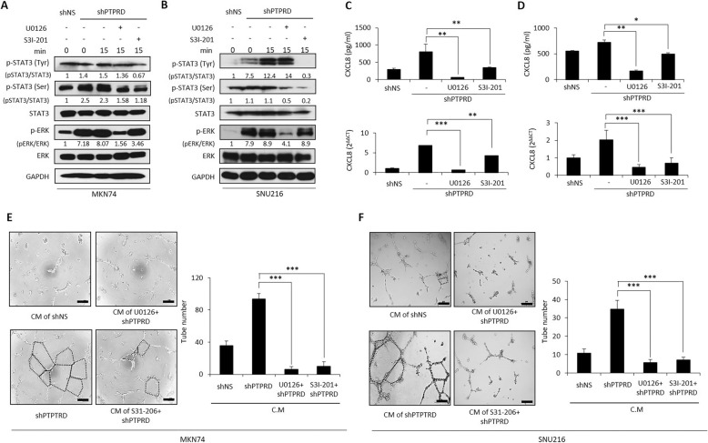
Fig. 4.
Loss of PTPRD activates ERK and STAT3 signaling. a-b Western blots after treatment of MKN74 and SNU216 cells with U0126 (ERK inhibitor) and S3I-201 (STAT3 inhibitor). Cells were collected after pretreatment with U0126 (10 μM) and S3I-201 (100 μM) for 4 h. c-d CXCL8 mRNA and secreted CXCL8 were measured by qRT-PCR and ELISA, respectively. Cancer cells were treated with each inhibitor for 48 h. e-f In vitro tube formation assay using MKN74 and SNU216 cells. Lentivirus-transfected cancer cells were treated with control (con) siRNA or CXCL8 siRNA for 48 h and the media were then changed to supplement-free HUVEC media. After 24 h, HUVECs were incubated in these conditioned media for 6–10 h. These data are representative of three independent experiments. *p < 0.05; **p < 0.01; ***p < 0.001 by unpaired Student’s t-test. Scale bar = 200 μm

