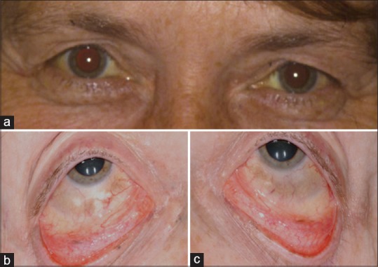
Figure 1.

Bilateral scleral melanoctosis. (a) External photograph demonstrating minimal periocular dermal pigmentation, but there was dark blue temporal fossa pigmentation and there was diffuse episcleral melanocytosis of the right (b) and the left (c) eyes
