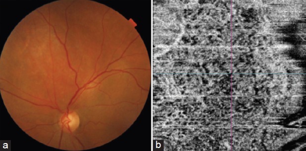
Figure 6.

(a) Fundus photograph of the left eye showing circumscribed choroidal hemangioma superior to the disc with subretinal fluid at the macula and (b) optical coherence tomography angiography showing a dense vascular network in the choriocapillary layer. (figure reprinted with permission from Dr Mahesh Shanmugam, Konana VK, Shanmugam PM, Ramanjulu R, Mishra KD, Sagar P. Optical coherence tomography angiography features of choroidal hemangioma. Indian journal of ophthalmology. 2018 Apr;66(4):581.)[28]
