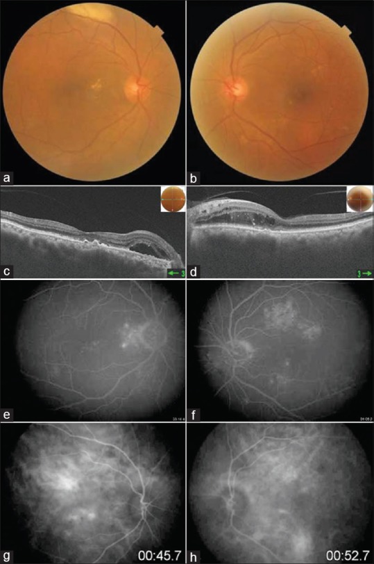
Figure 1.

Colour fundus photo shows multiple yellowish drusen like exudates with RPE alterations (a and b). OCT shows irregular RPE layer with presence of subretinal hyperreflective lesions and outer retinal edema (c and d). FA shows multiple hyperfluorescence areas withoutany leakage in late phase (e and f). ICG shows hypo and hypercyanscence spots with dilated choroidal vasculature (g and h)
