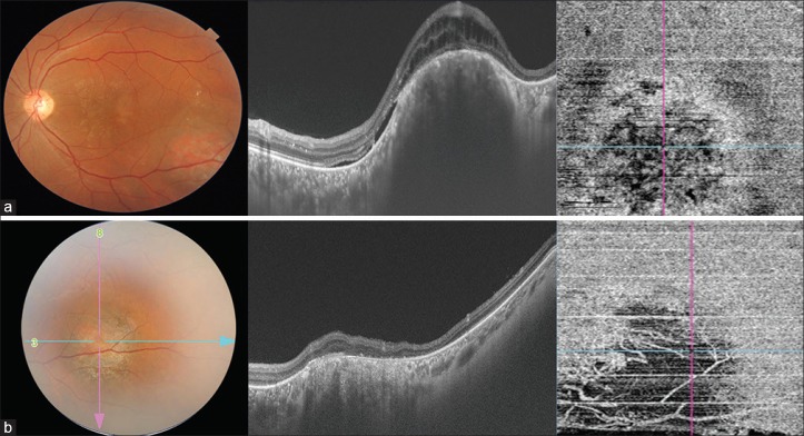
Figure 1.
(a) Fundus photograph – circumscribed orange-colored elevated lesion seen. OCT – dome-shaped choroidal hemangioma with presence of subretinal and intraretinal fluids over the tumor. OCT A in the choriocapillary layer – hyporeflective areas suggestive of loss of choriocapillaris with irregularly arranged vessels. (b) Fundus photograph post TTT – reduction in tumor size with RPE atrophy. OCT – decrease in tumor height and SRF and atrophy of outer retinal layers. OCT A showed complete loss of choriocapillaris extending beyond the margin of tumor and absence of deeper choroidal vasculature

