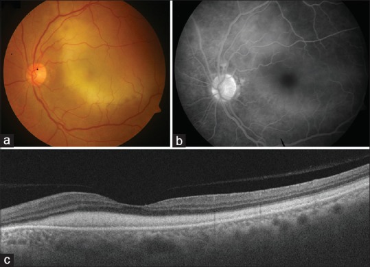
Figure 1.

Fundus photograph of the left eye of the patient described in the report showing presence of a large, diffuse yellowish submacular lesion (a). There is no evidence of any chorioretinal lesion. Fluorescein angiography (FA) shows presence of a diffuse hyperfluorescence in the macular region, corresponding to the yellowish submacular lesion (b). Swept-source optical coherence tomography shows presence of a diffuse sheet-like deposition of subretinal material in the macular region (c)
