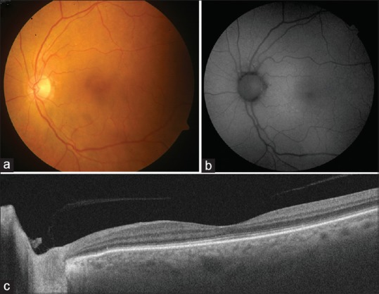
Figure 2.

Follow-up fundus photography of the left eye at 6 weeks after initial presentation showing complete resolution of the yellowish submacular lesion after initiation of intravitreal and systemic chemotherapy (a). Fundus autofluorescence imaging does not reveal any abnormal autofluorescence patterns indicating lack of retinal pigment epithelial damage (b). Follow-up swept-source optical coherence tomography shows resolution of the submacular deposits and normalization of foveal anatomy (c)
