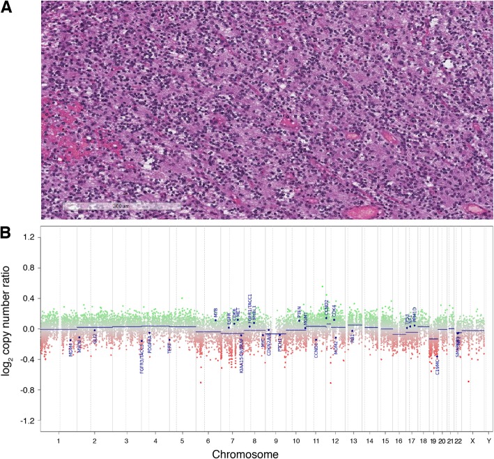
Fig. 2.
Histopathology and CNV plot for the tumor in Case 1 H&E stain and CNV plot for Case 1. a Hematoxylin and eosin stain of the tumor diagnosed as a Glioma, NOS with high-grade features and IDH1 (R132H) negative testing. b Relatively flat CNV plot supporting the integrated diagnosis of a dysembryoplastic neuro-epithelial tumor (DNET)

