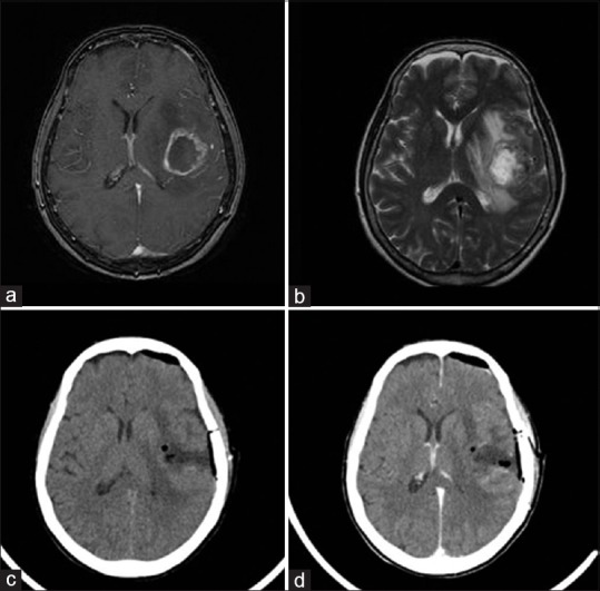
Figure 3.

(a-d) Case 2: (a) Axial contrast-enhanced T1-weighted magnetic resonance imaging demonstrating a heterogeneous cystic enhancing mass located intra-axially and in the extraventricular space in the left temporo-insular region. (b) Axial T2-weigheted magnetic resonance imaging demonstrating previous intratumoral hemorrhage. (c and d) Postoperative axial computed tomographic scan (c) and (d) contrast-enhanced computed tomographic scan demonstrating gross total resection
