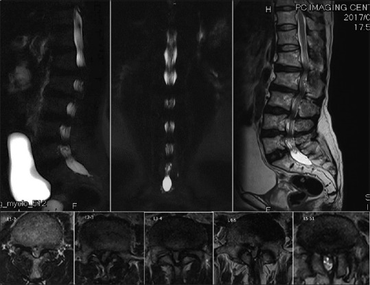
Figure 6.

Preoperative magnetic resonance imaging revealed markedly spinal canal stenosis from T10/11 and L1–L2 to L5/S1. The sagittal view showed multiple lumbar disc degeneration from L1/2 to L5/S1 with mild posterior bulging. The axial view revealed severe central and lateral recess stenosis
