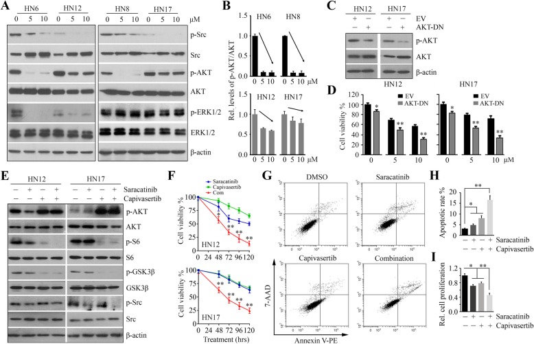
Fig. 2.
The addition of AKT inactivation augments the cytotoxicity of saracatinib in Sar-R HNSCC cells. a The effect of saracatinib on AKT and ERK1/2 signaling pathways in Sar-S (HN6 and HN8) and Sar-R (HN12 and HN17) HNSCC cells determined by Western blotting. b The quantitative data of phospho-AKT levels in saracatinib treatment from three independent experiments. c The effect of AKT-DN transfection on AKT activation determined by Western blotting. d The effect of AKT-DN transfection on cell viability in the presence or absence of saracatinib determined by CellTiter-Glo® Luminescent Cell Viability Kit on day 3 after treatment. e The effect of capivasertib on AKT-S6 signaling in the presence or absence of saracatinib determined by Western blotting. f The effect of 5 μM capivasertib on cell viability in the presence or absence of saracatinib determined by CellTiter-Glo® Luminescent Cell Viability Kit at 120 h after treatment. EV: empty vector; AKT-DN: a dominant-negative form of AKT (K179 M). g The effect of saracatinib, capivasertib and their combination on HN12 cell apoptosis determined on Day 3 after treatment using Annexin V: PE Apoptosis Detection Kit with 7-AAD. h The quantification of apoptotic rate from three independent experiments. i The effect of saracatinib, capivasertib and their combination on HN12 cell proliferation determined by MTS on Day 3 after treatment. *p < 0.05; **p < 0.01

