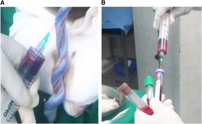
Figure 2.
Pictures taken during the study, illustrating the collection of umbilical cord blood samples. (A) Sampling 20 mL of cord blood with syringe. (B) Filling three vacutainer tubes with ∼ 6 mL each. This figure appears in color at www.ajtmh.org.

