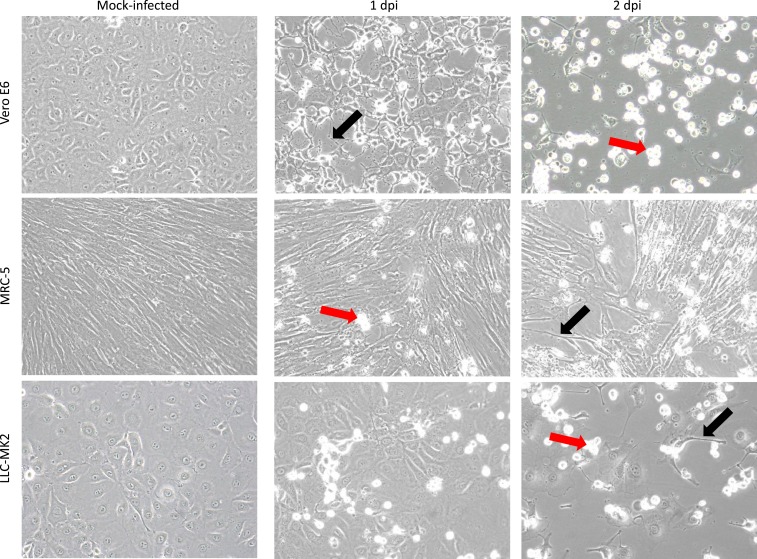
Figure 3.
Isolation of Mayaro virus in cell culture. Mock-infected Vero E6, MRC-5, and LLC-MK2 cells are shown 2 days postseed in the left panels. Cells inoculated with plasma from patient HI_112 are shown 1 dpi and 2 dpi in the middle and right panels, respectively. Refractile floating dead cells are evident (red arrows) and virus-induced cytopathic effects include apoptosis and appearance of cells with shrunken cytoplasm (black arrows). Original images at ×200 magnification. This figure appears in color at www.ajtmh.org.

