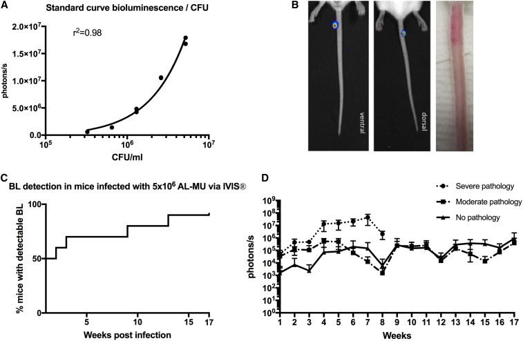
Figure 1.
Use of bioluminescent Mycobacterium ulcerans to follow the evolution of disease in the mouse tail model of Buruli ulcer. (A) Standard curve comparing bioluminescence and colony-forming units (CFUs) of the M. ulcerans JKD8049+pMV306 hsp16 luxG13 reporter strain. (B) IVIS (left) and photographic images (right) from BALB/c mice infected via subcutaneous tail inoculation with approx. 3.3 × 10⁵ CFU/mL M. ulcerans harboring the bioluminescent reporter plasmid. Photons/s emitted from bioluminescent bacteria were detected by IVIS in anesthetized mice. Results are represented in a pseudo-colored scheme (red indicated high, yellow indicated medium, and green indicated low intensity of the light emitted). Light was detected from both the dorsal (site of injection) and ventral aspects of the mouse tail. The photos were taken at 4 weeks postinfection; the bioluminescence readout was 1.8 × 10⁶ photons, corresponding to approx. 6.7 × 10⁵ CFU/mL according to our standard curve. (C) Survival graph representing time to detectable bioluminescence emission from mice infected with bioluminescent M. ulcerans into the tail (D) Development of mean photons/s emitted from mice infected with 3.3 × 10⁵ bioluminescent M. ulcerans into the upper third of the dorsal tail. Values represent the mean of dorsal and ventral photons/s measurement. Animals were subgrouped for analysis by clinical staging based on severity of the gross pathology (severe: redness, swelling, edema, and impending ulceration; moderate: redness and edema; and no pathology. This figure appears in color at www.ajtmh.org.

