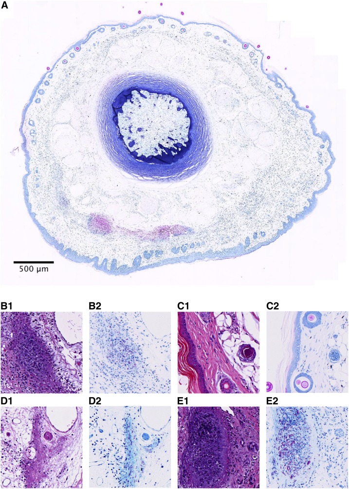
Figure 4.
Histological specimens of mice infected with Mycobacterium ulcerans into the tail in hematoxylin and eosin (A1, B1, C1, and D1) or Ziehl–Neelsen (A.2, B.2, C.2, and D.2) staining. (A) Whole slide cross-section of mouse tail (ID #85) infected with M. ulcerans subcutaneously, humanely killed 8 weeks postinfection because of advanced clinical pathology. Visible are clusters of acid-fast bacilli (AFB) and in the cutis and subcutis, approx. 300–400 μm beneath the surface (B.1 and B.2). Example of the presence of AFB in mouse 85, as well as granuloma formation (C.1 and C.2). Normal mouse tail histology of a naive, uninfected mouse with thin epidermis and intact hair follicles. (D.1 and D.2) Moderate pathology (mouse 89) with diffuse inflammation, tissue damage, and presence of AFB. Involvement of blood vessels with zones of inflammation and also destruction of smaller vessels was visible in this specimen too (vasculopathy) (E.1 and E.2). Necrosis, granuloma formation, inflammation, and abundant extracellular clustering of AFB were observed in mice with severe pathology (mouse 84). This figure appears in color at www.ajtmh.org.

