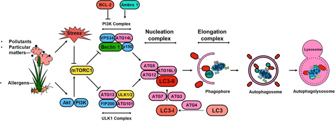
Figure 1.
Schematic overview of autophagy regulation. Environmental signals, such as environmental pollutants and allergens, induce cellular stress leading to the activation of the mTOR signaling complex 1 (mTORC1). Induction of autophagy begins with the formation of the phagophore, which is initiated by the ULK complex, consisting of ULK1 (or ULK2), autophagy-related protein 13 (ATG13), FAK family kinase interacting protein of 200 kDa (FIP200) and ATG101. PI3K complex, consisting of the vacuolar protein sorting 34 (VPS34) and the regulator subunits ATG14L, p150 and beclin 1, provides further nucleation signal. Autophagosome formation requires phagophore membrane elongation by a complex composed of ATG5, ATG12, ATG16L, and LC3-II, which are derived from the microtubule-associated protein 1 light chain 3 (LC3) by the activity of ATG4 generating LC3-I and the conjugation C-terminal glycine of LC3-I to phosphatidylethanolamine by ATG7, and ATG3. The formation of the autophagolysosome is a result of the fusion between the autophagosome and lysosomal compartments. Lysosomal hydrolyases degrade the autophagy cargo in all three processes.

