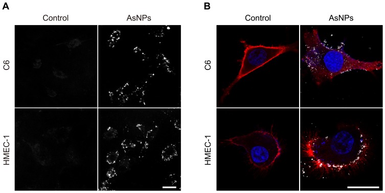
Figure 4.
Tumor-targeting property of AsNPs at the cellular level.
Notes: C6 glioma cells and normal human microvascular endothelial cells (HMEC-1) were used in the experiment. (A) Representative dark-field images. Scale bar: 25 μm. (B) Representative confocal images. The cell membranes were stained with DiD (red color), while the cell nuclei were stained with DAPI (blue color). The bright spots represented AsNPs. Scale bar: 25 μm.
Abbreviations: AsNPs, PEG- and As1411-functionalized silver nanoparticles; μm, micrometer; DiD, 1,1′-dioctadecyl-3,3,3′,3′-tetramethylindodicarbocyanine; DAPI, 4′,6-diamidino-2-phenylindole dihydrochloride.

