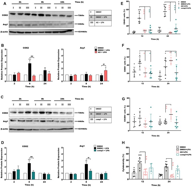
Figure 4.
AS2717638 and compound 3 restore a neuroprotective microglial phenotype while only AS2717638 reduces neurotoxicity of microglia-conditioned medium. Serum-starved microglia cells were treated with DMSO, DMSO plus LPA (1 μM), and LPA plus AS2717638 (0.1 μM; A) or compound3 (1 μM; C) for 2, 8, and 24 h. Cell lysates were collected and the expression of COX-2 and Arg-1 was monitored by immunoblotting. One representative plot for each protein and the densitometric analysis (B,D; mean ± SD) from three independent experiments are presented (mean value of DMSO controls was set to 1). In a parallel experiment, serum-starved (o/n) BV-2 cells were cultivated in the presence of DMSO, DMSO plus LPA (1 μM) or LPA in the presence of AS2717638 (0.1 μM) or compound 3 (1 μM) for the indicated times. Cells were stained with PE-conjugated anti-CD40 (E), APC-conjugated anti-CD86 (F) or PE-conjugated anti-CD206 (G) antibodies and analyzed using a Guava easyCyte 8 Millipore flow cytometer. Results are shown as mean values ± SD. (H) CATH.a neurons were incubated for 24 h with conditioned media collected from LPA-treated BV-2 cells in the presence or absence of AS2717638 (0.1 μM) or compound 3 (1 μM) for 12 and 24 h. The LDH levels were detected and cytotoxicity was calculated according to the manufacturer’s instructions (*p < 0.05; **p < 0.01; ***p < 0.001 compared to DMSO-treated cells; #p < 0.05; ##p < 0.01; ###p < 0.001 each inhibitor compared to LPA-treated cells; one-way ANOVA with Bonferroni correction).

